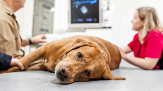
Caring for pets involves understanding their health needs at every stage of life. When a pet faces cancer, diagnostic imaging becomes crucial. Tools like X-rays, ultrasounds, and MRIs help vets pinpoint issues and plan treatment. In the field of veterinary oncology, these imaging methods offer a clear view of tumors and other concerns. For instance, MRI colorado is a trusted resource for veterinarians looking to gather precise images. Diagnostic imaging plays a vital role in building a clear path forward for our animal companions.
Understanding Diagnostic Imaging
Diagnostic imaging covers various techniques used to visualize the inside of a pet’s body. Each type of imaging serves a unique purpose. For example, X-rays are great for seeing bones. They help detect fractures or abnormalities. Ultrasounds, on the other hand, are used to view soft tissues. They are often used in pregnancy checks or to see organ changes.
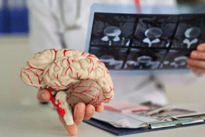
MRIs offer a deeper look. They provide detailed images of bones, organs, and tissues. This allows vets to see tumors more clearly. With this information, veterinarians can make informed decisions about treatment options, whether it involves surgery, chemotherapy, or other methods.
The Role of Imaging in Veterinary Oncology
In veterinary oncology, imaging plays a significant role in diagnosing and treating cancer in pets. Here’s how:
- Diagnosis: Imaging helps identify tumors early. Early detection can lead to better outcomes.
- Staging: Determines how far cancer has spread. Helps in planning treatment.
- Monitoring: Tracks the progress of treatment. Helps vets adjust plans if needed.
Comparing Imaging Techniques
Each diagnostic tool has its strengths. Below is a comparison table highlighting the key features of X-rays, ultrasounds, and MRIs:
| Imaging Technique | Best For | Limitations |
| X-rays | Bone fractures and abnormalities | Limited detail on soft tissues |
| Ultrasounds | Soft tissues and organ changes | Less effective for bone detail |
| MRIs | Detailed imaging of bones, organs, and tissues | Higher cost and longer time |
Advancements in Veterinary Imaging
Technological advancements have made imaging more accessible and effective. Vets now use digital X-rays for quicker results. Portable ultrasounds allow for on-the-go checks. Magnetic Resonance Imaging has improved, offering better resolution and clarity.
These advancements mean more accurate diagnoses and better outcomes for our pets. Vets can now detect diseases earlier and tailor treatments to individual needs. Early intervention often results in more effective treatment and a higher quality of life for pets.
Preparing Pets for Imaging
Before imaging, veterinarians offer guidance on preparing pets. This might include fasting for a few hours or calming strategies. Most pets handle the process well. Sedation is sometimes needed to keep them still, especially during MRI scans. The safety and comfort of pets during these procedures are always a top priority.
The Future of Veterinary Imaging
As technology advances, so does the potential for even greater innovations in veterinary imaging. Research continues to explore new techniques that could offer more detailed insights with less discomfort and cost. The use of artificial intelligence in analyzing images is one such area of exploration. This could lead to faster, more accurate diagnosis and treatment plans.
In summary, the role of diagnostic imaging in veterinary oncology cannot be overstated. It offers a window into the health of our pets, guiding us in making the best decisions for their care. With continued advancements, the future looks promising for even better diagnostic and treatment options.


