1. Introduction to HOCM
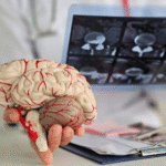
Hypertrophic obstructive cardiomyopathy (HOCM) is a genetic heart condition characterized by abnormal thickening of the heart muscle, particularly the left ventricle, leading to impaired cardiac function.
2. Diagnostic Challenges
Diagnosing HOCM can be challenging due to its varied clinical presentation and overlapping features with other cardiac conditions.
3. Role of ECG
Electrocardiography (ECG) plays a crucial role in the initial evaluation and diagnosis of HOCM, providing valuable insights into the electrical activity of the heart.
4. Typical ECG Findings
Common ECG findings in HOCM include left ventricular hypertrophy (LVH), repolarization abnormalities, and arrhythmias, which may aid in suspecting and confirming the diagnosis.
5. Left Ventricular Hypertrophy
LVH on ECG is characterized by increased QRS complex voltage in the precordial leads (V1-V6) and may exhibit repolarization abnormalities such as ST-segment depression or T wave inversion.
6. Asymmetric Septal Hypertrophy
Asymmetric septal hypertrophy, a hallmark feature of HOCM, may manifest as tall R waves in the precordial leads (V1-V3) with deep S waves in the lateral leads (I, aVL, V5-V6), known as the “strain pattern.”
7. Pseudo-Infarct Pattern
The presence of deep, symmetric T wave inversions in the precordial leads (V1-V4) without accompanying ST-segment elevation or Q waves may mimic an anterior myocardial infarction, termed the “pseudo-infarct pattern.”
8. Arrhythmias
HOCM patients are prone to various arrhythmias, including atrial fibrillation, ventricular tachycardia, and sudden cardiac death, which may be evident on ECG as irregular rhythms or widened QRS complexes.
9. Left Atrial Enlargement
Left atrial enlargement, secondary to elevated left ventricular pressures, may be reflected on ECG by prolonged P wave duration (>120 ms) and biphasic P waves in lead V1, suggestive of left atrial overload.
10. Atrial Fibrillation
Atrial fibrillation is a common arrhythmia in HOCM patients and may present on ECG as irregularly irregular R-R intervals with absent P waves and chaotic fibrillatory waves.
11. Ventricular Hypertrophy Patterns
Differentiating between HOCM and other causes of LVH, such as hypertension or aortic stenosis, requires careful analysis of ECG findings, including the presence of strain pattern and absence of LV strain.
12. Dynamic Obstruction
Dynamic left ventricular outflow tract (LVOT) obstruction, characteristic of HOCM, may manifest as changes in the ECG pattern during maneuvers that alter preload and afterload, such as the Valsalva maneuver or exercise.
13. Provocative Maneuvers
Performing provocative maneuvers during ECG acquisition, such as standing from a squatting position or administering amyl nitrite, can elicit changes in LVOT obstruction and help confirm the diagnosis of HOCM.
14. Risk Stratification
ECG findings, along with clinical parameters and imaging studies, aid in risk stratification and management decisions, including the need for implantable cardioverter-defibrillator (ICD) placement or septal reduction therapy.
15. Genetic Testing
Identifying specific genetic mutations associated with HOCM, such as mutations in the MYH7 or MYBPC3 genes, may complement ECG findings and provide valuable prognostic information for affected individuals and their families.
16. Long-Term Monitoring
Regular ECG monitoring is essential for assessing disease progression, monitoring arrhythmias, and evaluating treatment response in HOCM patients undergoing medical therapy or invasive interventions.
17. Multidisciplinary Approach
Managing HOCM requires a multidisciplinary approach involving cardiologists, genetic counselors, and other specialists to optimize patient care, genetic counseling, and family screening for inherited cardiac conditions.
18. Lifestyle Modifications
Encouraging lifestyle modifications, such as avoiding strenuous physical activity and maintaining a healthy weight, helps reduce the risk of exacerbating LVOT obstruction and adverse cardiovascular events in HOCM patients.
19. Medication Management
Medical therapy for HOCM aims to alleviate symptoms, prevent arrhythmias, and improve cardiac function through the use of beta-blockers, calcium channel blockers, and antiarrhythmic medications, tailored to individual patient needs.
20. Septal Reduction Therapies
For symptomatic HOCM patients refractory to medical therapy, septal reduction therapies such as septal myectomy or alcohol septal ablation may be considered to alleviate LVOT obstruction and improve symptoms.
21. Family Screening
Given the genetic nature of HOCM, screening family members of affected individuals for the presence of genetic mutations and cardiac abnormalities is crucial for early detection and intervention.
22. Prognosis
The prognosis of HOCM varies depending on disease severity, extent of LVOT obstruction, presence of arrhythmias, and response to treatment, with regular follow-up and monitoring essential for optimizing long-term outcomes.
23. Patient Education
Educating patients about the nature of their condition, potential complications, and the importance of adherence to treatment and follow-up care empowers them to actively participate in managing their health and well-being.
24. Advocacy and Support
Advocacy organizations and support groups provide valuable resources, information, and community support for individuals living with HOCM and their families, fostering awareness, education, and research initiatives.
25. Conclusion
ECG analysis plays a pivotal role in the diagnosis, risk stratification, and management of HOCM, providing valuable insights into cardiac morphology, arrhythmias, and dynamic LVOT obstruction. Integrating ECG findings with clinical assessment, genetic testing, and imaging studies enables comprehensive care and personalized treatment strategies for HOCM patients.


